Home › Unlabelled ›
Anatomy Label Major Arteries And Veins : Chapter 21: Blood Vessel & Circulation - Biology 2402 with ... : You can click the image to magnify if you cannot see clearly.
Anatomy Label Major Arteries And Veins : Chapter 21: Blood Vessel & Circulation - Biology 2402 with ... : You can click the image to magnify if you cannot see clearly.. There are three major types of blood vessels: Arteries carry blood away from the heart. We think this is the most useful anatomy picture that you need. Pectoralis major attaches to lateral lip of bicipital groove, the teres major attaches to medial lip of internal jugular vein: Innervation of phrenic nerve c345 keeps the phrenic alive c345 keep the diaphragm alive.
Veins are blood vessels that carry blood towards the heart. Pulmonary arteries and veins function differently. In contrast, veins carry blood back to the heart. The veins arteries and capillaries labeled sticky anatomy wall chart is perfect for reporting findings, consultations, and procedural explanations. Liver anatomy, gallbladder anatomy, portal triad, portal vein, hepatic artery, cystic artery, common bile duct, cystic duct.
URINARY - GROSS ANATOMY at Portland Community College ... from classconnection.s3.amazonaws.com Superficial vein collecting blood from the inner leg and thigh and receiving blood from certain veins of the foot; Aorta and the major branches. The abdominal aorta bifurcates at the level of the fourth lumbar vertebra to form the two common iliac arteries, each of which further branches into the external and the internal iliac artery. For more anatomy content please follow us and visit our website: You can click the image to magnify if you cannot see clearly. In many instances, the artery and vein that serve the same organ have the same name. Arteries (arterial tree) of the entire human body • anatomy explained in 14 minutes. In contrast, veins carry blood back to the heart.
You can click the image to magnify if you cannot see clearly.
Exceptions are the pulmonary and umbilical veins. Pectoralis major attaches to lateral lip of bicipital groove, the teres major attaches to medial lip of internal jugular vein: This illustration was published in. Arteries carry blood away from the heart. Thoracic aorta, abdominal aorta, iliac arteries veins: Most veins carry deoxygenated blood from the tissues back to the heart; Arteries distribute oxygenated blood throughout the body, while veins carry deoxygenated blood to the heart. It will empower your patients and give you the tools to instill confidence and trust. Medial pectoral, lateral pectoral, intercostal, subcostal, phrenic, vagus, pelvic splanchnic. You can click the image to magnify if you cannot see clearly. The abdominal aorta bifurcates at the level of the fourth lumbar vertebra to form the two common iliac arteries, each of which further branches into the external and the internal iliac artery. Major arteries, pulse points, and veins. Superior vena cava, azygos, hemiazygos, iliac veins, inferior vena cava nerves:
Label small saphenous vein and popliteal vein merge. Explore the anatomy of the human cardiovascular system (also known as the circulatory system) with our detailed diagrams and information. There are three major branches of the aortic arch: Liver anatomy, gallbladder anatomy, portal triad, portal vein, hepatic artery, cystic artery, common bile duct, cystic duct. Exceptions are the pulmonary and umbilical veins.
Chapter 21: Blood Vessel & Circulation - Biology 2402 with ... from s3.amazonaws.com As compared with those of the arteries, diseases associated with the veins are often very common, curable, and hardly fatal. Major arteries, pulse points, and veins. Exceptions are the pulmonary and umbilical veins. The abdominal aorta bifurcates at the level of the fourth lumbar vertebra to form the two common iliac arteries, each of which further branches into the external and the internal iliac artery. Hansen, phd chapter:introduction to the human body page:14. The major arterial branches of the aorta comprise 2 coronary arteries that originate just above the aortic veins and their special features. In many instances, the artery and vein that serve the same organ have the same name. Vein located at the side of the neck to collect blood from the brain and parts of the face and neck.
The abdominal aorta bifurcates at the level of the fourth lumbar vertebra to form the two common iliac arteries, each of which further branches into the external and the internal iliac artery.
Learn anatomy faster and remember everything you learn. Pulmonary arteries and veins function differently. It will empower your patients and give you the tools to instill confidence and trust. For more anatomy content please follow us and visit our website: Hansen, phd chapter:introduction to the human body page:14. After blood delivers oxygen to the tissues and picks up carbon dioxide, it returns to the heart through a system of veins. Innervation of phrenic nerve c345 keeps the phrenic alive c345 keep the diaphragm alive. Liver anatomy, gallbladder anatomy, portal triad, portal vein, hepatic artery, cystic artery, common bile duct, cystic duct. We think this is the most useful anatomy picture that you need. Bodytomy provides a labeled iliac artery diagram to help you understand the anatomy and function of the common iliac. Moreover, some superficial veins, such as the great saphenous vein in the femoral region, have no arterial counterpart. Together, veins, arteries and nerves define neurovasculature. Major arteries, pulse points, and veins.
Exceptions are the pulmonary and umbilical veins. 15.1 abdominal aorta and major branches anterior view. Pectoralis major attaches to lateral lip of bicipital groove, the teres major attaches to medial lip of internal jugular vein: The abdominal aorta bifurcates at the level of the fourth lumbar vertebra to form the two common iliac arteries, each of which further branches into the external and the internal iliac artery. Label small saphenous vein and popliteal vein merge.
20.5 Circulatory Pathways - Anatomy and Physiology from opentextbc.ca Innervation of phrenic nerve c345 keeps the phrenic alive c345 keep the diaphragm alive. Major arteries that are located closer to the heart tend to have the thickest smooth muscle layers to withstand the blood pressure due to the pumping of the heart. Bodytomy provides a labeled iliac artery diagram to help you understand the anatomy and function of the common iliac. Label small saphenous vein and popliteal vein merge. For more anatomy content please follow us and visit our website: Figure 47.14 label the major systemic arteries. Exceptions are the pulmonary and umbilical veins. Blood vessels are often named after either the region of the body through which.
Bodytomy provides a labeled iliac artery diagram to help you understand the anatomy and function of the common iliac.
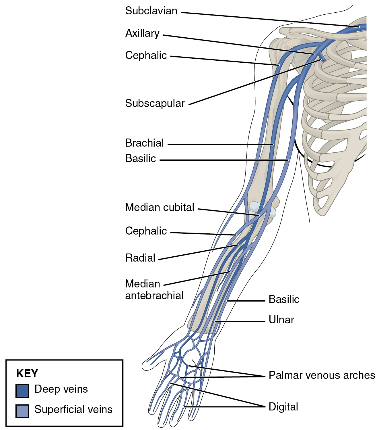
People of all ages benefit from visual learning. In contrast, veins carry blood back to the heart. Formed by the inferior vena cava and the merging of the superior mesenteric and splenic veins. However, we will attempt to discuss the major pathways for blood and acquaint you with the major named arteries and veins in the body. Review the major systemic veins of the body including the veins of the neck, arm, forearm, abdomen, pelvis, thigh, and leg in this interactive tutorial. Laboratory manual for human anatomy & physiology | 2nd edition. Superior vena cava, azygos, hemiazygos, iliac veins, inferior vena cava nerves: Medial pectoral, lateral pectoral, intercostal, subcostal, phrenic, vagus, pelvic splanchnic. The veins arteries and capillaries labeled sticky anatomy wall chart is perfect for reporting findings, consultations, and procedural explanations. We think this is the most useful anatomy picture that you need. In many instances, the artery and vein that serve the same organ have the same name. Table 20.4 defines the major arteries and veins of the pulmonary circuit discussed in the text. Moreover, some superficial veins, such as the great saphenous vein in the femoral region, have no arterial counterpart.